1. What's the purpose of the pericardium?
- Surround and protect the heart
- Fibrous pericardium anchors the heart and provides protection through its tough, inelastic, and dense irregular tissue
2. Observe the blood vessels connecting to the heart. How do arteries differ from veins in their structure?
- Arteries have thicker walls and veins have thinner walls
3. What function do you think the auricle serves?
- It expands the blood-holding capacity of atrium
4. What differences do you observe between the atria and ventricles?
- The atria are smaller than the ventricles and the ventricles collect and expel blood from the atria into the body or the lungs
5. Use words or pictures to describe each
- Coronary sinus
- blood vessel that goes across the back of the heart
B. Inferior vena cava
- doesn’t have it because it was cut too close
C. Tricuspid valve
- looks like a flap
6. Draw a picture of tricuspid valve, including chordae tendinae and the papillary muscle
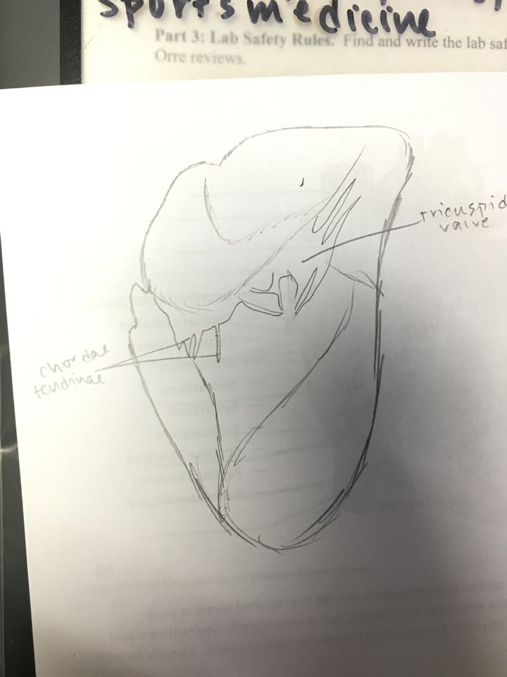
7. Why is the “anchoring” of the heart valves important?
- It’s important so that the valve doesn’t get carried away with the blood
8. Use words and pictures to describe what you see.
9. What’s the function of the semilunar valve?
- It prevents blood from flowing back into the heart
10. If the valve disease occurs on the right side of the heart, it results in swelling in the feet and ankles.
(a) Why might this happen?
(a) Why might this happen?
- Blood is supposed to leave the feet and ankles and go through the right side of the heart, but with this disease, there is a backflow of blood because the valves are improperly functioning
(b) If the valve disease occurs on the left side of the heart, what complications would you expect to see?
- You would see that you aren’t getting a sufficient enough of blood to the muscles and body and therefore you would notice you have shortness in breath
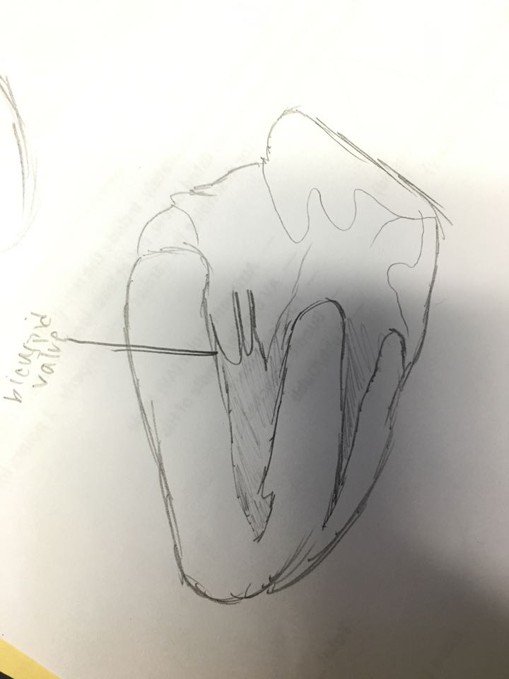
11. Using pictures and/or words, describe what you see.
(a) Entrance to the right and left coronary arteries: they connect to the aorta and they revolve around the back of the heart
(b) Left aortic semi-lunar valve: looks like a flap
(c) Chordae tendinae of the bicuspid valve: fibrous and tendon-like chords that connect the valve to the papillary muscle
(d) Papillary muscle of the biscupid valve: connects to the bottom of the left ventricle, lump of muscle that connects to the chordae tendinae and helps open and close the bicuspid valve
12. Describe how the left and right sides of the heart differ from each other.
- left side is more muscular → pumps blood through body
- right side is not as muscular because it pumps blood to lungs
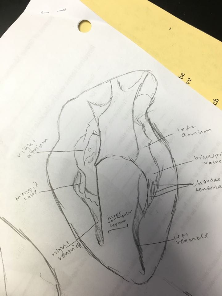
13. Draw and label all structures visible in the interior of the cross-section
No comments:
Post a Comment